**Disclaimer - This post contains images of the skull. View at your own discretion**
When we adopted Molly nine years ago, we knew that she would have some surgery but based on the little information that we had at the time and the fact that she already had a surgery in China prior to us adopting her, we naively believed that her surgery would be purely cosmetic, to improve the appearance of the scarring that existed on her face.

We had no idea that there was anything that needed to be corrected "on the inside". We were shocked and even frightened when her first CT scan revealed a fairly significantly sized hole in between her eyes.

Molly had to have a surgery to correct both the hole left by her encephalocele and her hypertelorism, which was the widened space between her eyes. The surgery is known as "facial bi-partition", but is similar to a "box osteotomy" based on the wikipedia explanation below (easier to copy/paste than type in my own words).


Box osteotomy
This treatment of orbital hypertelorism was first performed by Paul Tessier.[4] The surgery starts off by various osteotomies that separate the entire bony part of the orbit from the skull and surrounding facial bones. One of the osteotomies consists of removing the bone between the orbits. The orbits are then mobilized and brought towards each other. Because this often creates excessive skin between the orbits a midline excision of skin is frequently necessary. This approximates the eyebrows and eye corners and provides a more pleasing look.[1]
Facial bipartition
The standard procedure (box osteotomy) was modified by Jacques van der Meulen and resulted in the development of the facial bipartition(or median faciotomy).[5][6] Facial bipartition first involves splitting the frontal bone from the supraorbital rim. Then the orbits and the midface are released from the skull-base using monoblock osteotomy. Then a triangular shaped piece of bone is removed from the midline of the midface. The base of this triangular segment lies above the orbita and the apex lies between the upper incisor teeth. After removing this segment it is possible to rotate the two halves of the midface towards each other, thus resulting in reduction of the distance between the orbits. It also results in leveling out the V-shaped maxilla and therefore widening of it. Because hypertelorism is often associated with syndromes like Apert, hypertelorism is often seen in combination with midfacedysplasia. If this is the case, facial bipartition can be combined with distraction osteogenesis. The aim of distraction osteogenesis of the midface is to normalize the relationship between orbital rim to eye and also normalize the position of zygomas, nose and maxilla in relation to the mandible.[7]

The photos below show Molly's skull six weeks after her first surgery.
This inside of her face, including her eyes and palate was shifted.


The section of bone that was removed from the front of Molly's skull, the portion that contained the hole, was "recycled" to fill the gap produced by the shift.

After 15 months, the gap was nearly completely filled in. Bone will regenerate and using a person's own bone for grafting is better as it is the same DNA working together so it results in a quicker and stronger graft as opposed to using animal bone, for example.

This is Brynn's skull prior to her first surgery. As you can see, it is nearly perfect with the exception of the "notch" of bone missing on the right side of her nasal cavity.


I'm showing these photos and giving these explanations related to the girls' medical conditions when we adopted them so that you can realize with us the magnitude of difference and severity when compared to the boys' scans that were done at the beginning of this week.

Once again, based on our experience with Molly and Brynn, we felt that we were well equipped for Jonah and Rees' medical situation and what would need to be done for them. But, once again, we are again surprised by unexpected information.
I am showing this photo of Molly to give you a normal perspective on the jaws and the adult teeth that have not yet come through. Despite Molly's medical condition, her jaws and her teeth were unaffected.

The scan below is of Jonah's skull. The first one is the transparency, similar to the one of Molly above. However, you can see that in the area of Jonah's baby teeth, as well as his adult teeth, the white objects that show his teeth are jumbled and jagged. Not one of his adult teeth appear to be in the right (normal) place.

So, this is Jonah's skull. You might not notice it being too different at first look, which is why I provided the above photos for comparison.
Jonah was born with a very rare and serious facial cleft. According to the volunteer surgical team that performed the first surgery that Jonah had earlier this year in China, the cleft was a Bilateral Tessier Facial Cleft #7.
We, of course, told our very experienced, well-known, super-duper brilliant, Professor of a Surgeon and he said something we were not expecting ...
He said that he has NEVER seen anything like this!

Jonah's cleft was most likely caused by amniotic banding but this is just the highest assumption. The banding was very severe and went completely across his mouth and face.


The band was in place before the skull was still forming as the skull did it's best to form around it creating the malformations in various parts of the bone and flesh.


Jonah's lower jaw is at least an inch and a half recessed due to the cleft and his palate is extremely short. The left side of his face was affected more severely. His cheek bones are not connected as the band went right through the area that those bones would be ... you can see the gap in the photo below.

The back and sides of the skull seem slightly enlarged and mishapen, which the surgeon did agree with, however, the other issues are obviously the main concern.
The next step is to see a special orthodontist in about a week and a half. She will evaluate whether or not Jonah should have "Jaw Distraction" immediately OR have cranial surgery to correct some of the malformations of his skull. This orthodontist is a colleague of the craniofacial surgeon so they will determine together the best plan. More to come ...

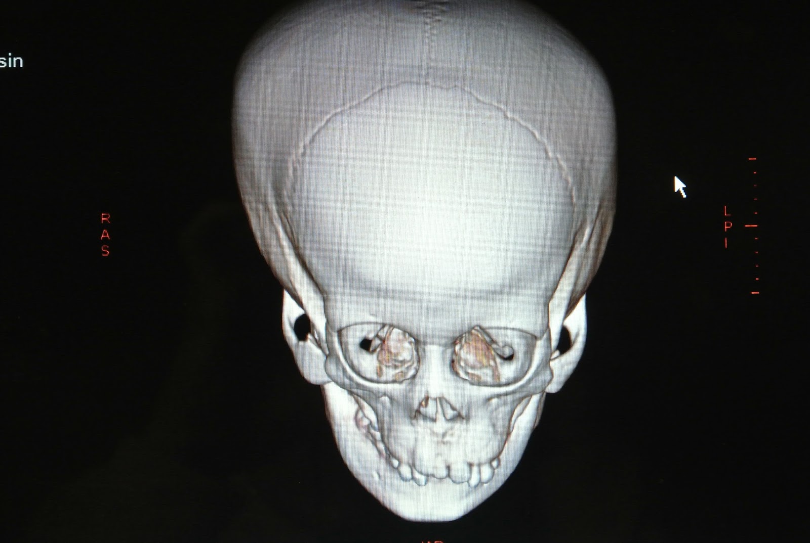
And then there's our little spit-fire, REES!!
Just look at his darling and amazing skull!

Rees' skull is NOT AT ALL what we expected!
Due to the size of the gap between his nose and distance between his eyes, we expected his scan to reveal an enormous hole ... like Molly's but BIGGER.

Do you see that teeny tiny hole in the center of the forehead shown in the photo below?
That is where the encephalocele is. That's how small of an opening it made!
Yet, it completely obliterated the formation of his nose!!
He may have had a nasal cavity at one point, however, his bone just kept forming and completely filled in behind the encephalocele, which is hanging downward out of the teeny-tiny opening and resting in that recessed area of bone between his nostrils.

You can also see where a cleft was starting in the portion of the skull that would have potentially affected the soft tissues such as the palate, however, his palate is unaffected. He has a small hole above the upper jaw contains a "floater" tooth and his upper lip has ever-so-slight evidence of the cleft but the main issue is his nose ...

... or lack of ...

While we can't necessarily see it by looking at these images, the surgeon pointed out that Rees has two nasal septums and two "something-or-other" nerves ... and two "this-or-thats" ... he said that it was as if the encepalocele attempted to cause the formation of two noses instead of one.


Rees is scheduled to have surgery in January and he will initially have the same surgery that Molly had. The surgeon did tell us that Rees' condition is much more severe than Molly's and that we should expect his treatment to go in stages.
We will share more about the surgery as it gets closer. In the meantime, we are going to try to enjoy December and Christmas and spending time together as we continue to bond with and get to know these two precious boys.
Today, they dipped marshmallows into dark chocolate and then added sprinkles.

They really enjoyed doing this ... and, of course, tasting them!

They enjoy getting outside into the fresh air ...

... and snuggling up in warm, footy pajamas!!

They like showing Mama their newest creation ...

... and playing airplane with Baba!!

Four weeks in the USA under our belt!
We're doing pretty well ... each day shows rays of hope and steps forward and the boys are really beginning to cling to our family routine.
We thank the LORD for bringing us this far, for holding onto us even when we felt like we were dangling and we thank the LORD for what He is yet to do in and through the lives of these boys.
Wow! I enjoyed reading this. It made me feel as though these boys are in the family they were meant to be in since birth. I wonder why it takes so long to find the home where they belong. Beautiful blog!
ReplyDelete